Day 2 :
Keynote Forum
Peter A. DePergola II
Director of Clinical Ethics, Baystate Health; Research Scientist in Neuroethics, American Academy of Neurology USA
Keynote: Contemporary Issues in Neuroethics
Time : 9:00 am

Biography:
Abstract:
Keynote Forum
Wai Kwong TANG
Chinese University of Hong Kong , China
Keynote: Structural and functional MRI correlates of Poststroke Depression
Time : 9:45 am

Biography:
Professor WK Tang was appointed to professor in the Department of Psychiatry, the Chinese University of Hong Kong in 2011. His main research areas are Addictions and Neuropsychiatry in Stroke. Professor Tang has published over 100 papers in renowned journals, and has also contributed to the peer review of 40 journals. He has secured over 20 major competitive research grants, including Health and Medical Research Fund, reference number: 02130726. Health and Medical Research Fund, reference number: 01120376. National Natural Science Foundation of China, reference number: 81371460. General Research Fund, reference number: 474513. General Research Fund, reference number: 473712. He has served the editorial boards of five scientific journals. He was also a recipient of the Young Researcher Award in 2007, awarded by the Chinese University of Hong Kong.
Abstract:
- Autonomic Neurology |Neurodegenerative Disorder | Recent Research| Neurobiology
Location: Conference Hall
Session Introduction
Stefan M. Brudzynski
Professor Psychology and Neuroscience Brock University,Canada Canada
Title: The Ascending Reticular Systems For Emotional Arousal

Biography:
Stefan M. Brudzynski, Ph.D., D.Sc., is a neurophysiologist, neuroscientist, and biopsychologist, presently Professor of Psychology and Neuroscience at Brock University, St. Catharines, ON, Canada. He is a former Director of the Centre of Neuroscience and the recipient of the prestigious Life-time Achievement Award from the International Behavioral Neuroscience Society for his contribution to the field of Behavioral Neuroscience. His main research interest is in the neural substrate of animal behavior, neuropsychopharmacology, animal vocal communication, and particularly, central control of ultrasonic vocalization and communication in rodents. His current research is focused on vocal expression of emotional states and brain systems for emotional arousal. He is editor of two fundamental handbooks on neural control of vocalization, Handbook of Mammalian Vocalization – An Integrative neuroscience Approach (2010), and Handbook of Ultrasonic Vocalization – A Window into the Emotional Brain (2018).
Abstract:
Functions of the reticular activating system are fundamental for maintenance of state of wakefulness, consciousness and vigilance. Evidences have cumulated suggesting that the activating system contains a group of several reticular systems that are specialized in many aspects of arousal. This talk will be focused on emotional arousal and will present two specialized emotional arousal systems that are working in parallel to the cognitive arousal system. In animals, emotional arousal is overtly signaled by emission of state-specific vocalizations that allow for qualitative and quantitative studies of the emotional process. Laboratory rat represents the best studied mammalian species and rat ultrasonic vocalizations have been studied in depths over the last 20 years. They can be divided into two main categories of calls expressing negative or positive emotional arousal and state. The valence-specific vocalizations are labeled for simplicity according to their usual sound frequency as 22 kHz vocalizations expressing aversive (negative) states as anxiety, discomfort and displeasure, while 50 kHz vocalizations expressing appetitive (positive) hedonic states as reward or its anticipation, and play joy. Based on the studies of brain control of ultrasonic vocalization emission, two arousal systems will be presented: (a) the ascending mesolimbic cholinergic system for aversive arousal and (b) the ascending mesolimbic dopaminergic system for appetitive arousal. These two ascending systems originate from tegmentum and their activity is based on two different neurotransmitters. Positive emotional state is induced by release of dopamine while negative emotional state is induced by release of acetylcholine. These transmitters are released in vast subcortical limbic regions and are not targeted at the neocortex as the cognitive arousal system is. Functions of these two tegmental systems are extremely fast and seem to depend on functionally antagonistic relationship between them that allows for development of positive or negative emotional arousal but not both these states together. Results of these studies are relevant to neuropsychiatry and understanding of emotional states, their disturbances, and their relationship to cognitive functions of the brain.
Emmanuel Brouillet
Head, Neurodegenerative Diseases Laboratory Molecular Imaging Research Center (MIRCen) DRF, CEA France
Title: Topic can not be disclosed before conference presentation (Speaker reccommendation)

Biography:
After a PhD in Neuropharmacology at Paris University, Dr Brouillet received post-doctoral fellowships to study Huntington’s disease pathogenesis at the Neurology Department of the Mass. General Hospita (Harvard Medical School) where he worked with Professor M. Flint Beal. He competed for and obtained a position at the renowned French research organization C.N.R.S in 1993. Since, he dedicated his career to tackle key issues related to neurodegenerative diseases including Huntington’s, Parkinson’s and Alzheimer’s diseases. He also launched research program on new experimental therapeutics and the development of novel brain imaging methods. Dr Brouillet is now Director of Research at C.N.R.S. and is Head of the Neurodegenerative diseases Lab at C.E.A. near Paris. He is the co-author of more than 130 peer-reviewed publications. He teaches Neurosciences at Paris University and also member of many local and international scientific committees and boards for governmental organizations and charities dedicated to patients with neurodegenerative diseases.
Abstract:
Marcio R.R. Cunha Ramalho
Senior Instructor The Clinic Trauma Center, Brazil
Title: Lumbar Angiolipoma Removal Via Percutaneos Endoscopic Interlaminar Approach

Biography:
Dr. MARCIO ROBERTTI RAMALHO DA CUNHA is senior instructor at The Clinic Trauma Centre at Brazil. He completed this medical schooling from Federal University of Rio Grande Do Norte in 1983 – 1989. After completing his medical schooling he worked as Medical Residence at Base hospital Brazil in year 1990 to 1993. He complete post-graduation course from R.W.T.H Hospital, Aachen-Germany( 1994) and Freie University,Berlim, Germany(1997).
He is titular member of Brazilian Neurosurgery Society (1994), Brazilian Spine Society (2009), Brazilian M.I.S Society (2012) and Latino American Study Group Of Neuroendoscopy / Glen (2016).
He holded the position of U.F.R.N, Assistant Professor Of The Neurosurgery Department ( 1996-1999 ),Senior Staff Of The Spinal Surgery Area Of Clinic Trauma Center /Natal-Rn (2007), Chairman Of Neurosurgery Service Of The General Hospital / Natal-Rn (2014) and Coordinator Of The Spinal Endoscopy Surgery Field - Ab Health Institute (2016)
Abstract:
Percutaneous endoscopic technique has been used to treat disk herniation and spinal stenosis ( 5,8,10 ), so far we have very few reports to treat benign spinal tumors under this minimally invasive treatment( 3 , 5 ) . We would like to present a single case of lumbar epidural angiolipoma removed using full endoscopic interlaminar approach as a safe and effective option to treat benign extra dural spinal tumor .We describe a 54 year-old man with a 3-month history of a progressively worsening low back pain without any other neurological signs.A magnetic ressonance demonstrated a dorsally located L1-L2 epidural lesion . We've performed a total removed under the basic steps of the Interlaminar approach . After opening the ligamentun flavum , the tumor was totally removed piecemeal under endoscopic guidance , The procedure lasted less than three hours with no support in intensive care unit and pathological examination confirmed extradural lumbar angiolipoma .he had a hospital discharge less than twelve hours after the surgical procedure with no neurological signs and using minor pain killer to control the back pain .E ven though we don´t have so much papers about this surgical practice, we think that is a feasible treatment and for sure we need more adapted endoscopic tools to accelerate the surgical time .
Joëlle Chabry
Université Côte d'Azur, Institute of Molecular and Cellular Pharmacology, France
Title: Potential therapeutic of the adipokine Adiponectin as antidepressant

Biography:
Abstract:
Epidemiological data indicate high comorbidity between metabolic and psychiatric disorders, particularly obesity and depression. Using a well-characterized anxiety/depressive-like mouse model consisting of continuous input of corticosterone for several consecutive weeks, we investigate the metabolic changes including weight, adiposity and plasma biological parameters (lipids, adipokines, and cytokines). In parallel, a panel of reliable behavioral tests was conducted to assessing numerous facets of the depression-like state, including anxiety, resignation, reduced motivation, loss of pleasure and social withdrawal. Our data show that chronic administration of corticosterone induced the parallel onset of metabolic and behavioral dysfunctions in mice. Potent adiponectin receptor agonists, prevented the corticosterone-induced early onset of moderate obesity and metabolic syndromes as well as successfully reversed the depression-like state. The possible mechanisms of actions of adiponectin receptor agonists have been deeply investigate as well as the routes of delivery, the dose-response and the side effects in three different mouse models of anxiety/depression-like. Our study highlights the pivotal role of the adiponergic system in the development of both metabolic and psychiatric disorders. Because about one third of depressive patients are resistant to currently available antidepressants, Adiponectin receptor agonists may constitute a novel and promising class of antidepressants.
- Sessions: Neurophysiology | Recent Research| Neural Engineering
Location: Conference Hall
Session Introduction
Ikeshima-Kataoka
Keio University School of Medicine, Japan
Title: Reactive Astrocyte has a Neuroimmunological Function in injured Mouse Brain

Biography:
Dr. Ikeshima-Kataoka was graduated from Keio University School of Medicine (Department of Microbiology) and got Ph.D. on the functional analysis of three calmodulin genes using transgenic mice. At the National Institute of Neuroscience as a postdoctoral fellow, researched on the molecular mechanism of neuronal development using fly genetics. Then, promoted back to Keio University School of Medicine (Department of Neuroanatomy) and started to focus on to the “reactive astrocytes” in injured mouse brain. At Jikei University School of Medicine, joined to immunological team in Institute of DNA Medicine and focus on to neuroimmunological analysis in injured brain using knockout mice. Then, promoted back to Keio University School of Medicine (Department of Pharmacology and Neuroscience) and found that some important molecules are concerned in neuroimmunological functions around injury site of the brain using microarray analysis. Now, using in vivo imaging system with live mouse to analyze functional role of “reactive astrocytes” around injured brain at Waseda University, Faculty of Science and Engineering.
Abstract:
In the central nervous system (CNS), some glial cells such as astrocytes and microglial cells become active to proliferate and secrete some of the inflammatory cytokines around lesion site when traumatic injury or inflammation occurred. However, it is still unclear for the functional role of those activated glial cells in the brain. To focus onto the regeneration of the CNS, we have been to analyze functional role of reactive glial cells in the brain with stab wound injury. As a brain injury model, stab wound using 27G needle was made on the mouse cerebral cortex from caudal to rostral axis. In the reactive astrocytes, one of the extracellular matrix molecule, tenascin-C (TN-C) is highly expressed, we analyzed TN-C functional role using TN-C-deficient mice (TN-C/KO) and found that TN-C is required for activation of astrocytes such as proliferating and induce secretion of some of the inflammatory cytokines in the injured brain and in the primary culture of astrocytes. Furthermore, TN-C has an important role for recovery of blood brain barrier (BBB) breakdown caused by stab wound in the brain. From these results, reactivation of astrocytes in the injured brain might have a functional role for BBB recovery in the CNS. On the other hand, since one of the water channels in the CNS aquaporin 4 (AQP4) is upregulated in activated astrocytes after the stab wound, we have analyzed AQP4 functional role in reactive astrocytes with injured brain using AQP4-deficient mice (AQP4/KO) compared to the wild type mouse (WT) brain. To label the proliferating cells, bromo-deoxy-uridine (BrdU) was used and that could be incorporated to the mice through the drinking water. Immuno-fluorescent staining analysis was performed on cardiac perfused mouse brain sections with
Sundas Hira
Lecturer at Riphah International University, Pakistan
Title: Antioxidants Attenuate Isolation- and L-DOPA-Induced Aggression in Mice
Biography:
Sundas Hira is working as a lecturer at Riphah Institute of Pharmaceutical Sciences, Riphah International University. She is contributing dedicatedly her best part in research and publications. She has her expertise in assessing the use or effectiveness of natural substances in neurodegenerative diseases. Her research work based on evaluating the effectiveness of antioxidants in treating the aggression explores the new pathways for preventing many crimes in society. Social crime occur due to the negative traits in personality such as aggression etc. This research probe the use of natural supplements in various disease resulting due to oxidative stress.
Abstract:
Aggression is a highly conserved and destructive behavior with intention to cause harm. Aggression is associated with negative traits such as choler, impulsivity, perturbation and proneness to violence. Antioxidants have been used for the treatment of many ailments for many years. The present study was carried out on Swiss male albino mice to explore the role of antioxidants at two dose levels, such as ascorbic acid (15.42 and 30.84 mg/kg), beta carotene (1.02 and 2.05 mg/kg), vitamin E (2.5 and 5.0 mg/kg), and N-acetyl cysteine (102.85 and 205.70 mg/kg) in the treatment of aggression. Aggression was induced by putting mice into artificial situations (isolating animals for one month and administration of L-DOPA). Swiss male albino mice (n = 330) were divided into 11 groups (Group I-control, group II-diseased, group III-standard group, group IV–V treated with ascorbic acid , group VI–VII treated with beta carotene, group VIII–IX treated with vitamin E, group X–XI treated with N-acetyl cysteine for 14 consecutive days). Endogenous antioxidants (glutathione, superoxide dismutase, and catalase) were analyzed to determine the antioxidant potential in oxidative stress. In isolated animals, high dose of vitamin E (5.0 mg/kg) was found to be more potent to placate the mice while all other antioxidants produced dose-dependent anti-aggressive effect except N-acetyl cysteine which had pronounced anti-aggressive effect at low dose (102.75 mg/kg). In acute anti-aggressive activity against L-DOPA induced aggression, low doses of vitamin E (2.5 mg/kg) and N-acetyl cysteine (102.75 mg/kg) and high dose of beta carotene (2.05 mg/kg) were effective to avert all aggression parameters. However, all test antioxidants were equally effective in chronic anti-aggressive studies against L-DOPA induced aggression. It may be concluded that selected antioxidants can alleviate the aggression which is a prodrome of many neurological disorder such as schizophrenia etc.
- Sessions: Neurodegenerative Disorder| Neurosurgery | Neuro - imaging
Location: Conference Hall
Session Introduction
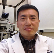
Peijun Li
Principal Investigator Second Affiliated Hospital Wenzhou Medical University Wenzhou, China
Title: Loss of CLOCK results in dysfunction of brain circuits that underlies pediatric focal epilepsy
Biography:
Dr. Peijun Li received his PhD from Department of Physiology and Biophysics, Georgetown University Medical Center. His research during his PhD study has been focusing on the inhibitory interneurons in mouse somatosensory cortex. During his postdoctorial work at Children’s National Medical Center at DC, he studied how hypoxia could affect the brain development using mouse model, and also how disruption in circadian gene expression could result in epilepsy in both human and mouse model. Dr. Li is now a principle investigator at Second Affiliated Hospital, Wenzhou Medical University, China. As a scientist with expertise in electrophysiology, his work will be directed towards understanding the functional roles of specialized GABAergic inhibitory circuits in the pathophysiological process of epilepsy and circuit formation. In his free time, Dr. Li likes running and listening to music. He have finished four half marathons and three full marathons which include the Marine Corps Marathon 2016 and the Rock and Roll DC 2016, His personal best is 3:53:25.
Abstract:
Because molecular mechanisms underlying refractory focal epilepsy are poorly defined, we performed transcriptome analysis on human epileptogenic tissue. Compared with controls, expression of Circadian Locomotor Output Cycles Kaput (CLOCK) is decreased in epileptogenic tissue. To define the function of CLOCK we generated and tested the Emx-Cre; Clockflox/flox and PV-Cre; Clockflox/flox mouse lines with targeted deletions of the Clock gene in excitatory and parvalbumin (PV)-expressing inhibitory neurons, respectively. The Emx-Cre; Clockflox/flox mouse line alone has decreased seizure thresholds, but no laminar or dendritic defects in the cortex. However, excitatory neurons from the Emx-Cre; Clockflox/flox mouse have spontaneous epileptiform discharges. Both neurons from Emx-Cre; Clockflox/flox mouse and human epileptogenic tissue exhibit decreased spontaneous inhibitory post-synaptic currents. Finally, video-EEG of Emx-Cre; Clockflox/flox mice reveals epileptiform discharges during sleep and also seizures arising from sleep. Altogether, these data show disruption of CLOCK alters cortical circuits and may lead to generation of focal epilepsy, which suggest that the circadian pathway may be a promising target for therapeutic intervention.
Marcio R.R. Cunha Ramalho
Senior Instructor The Clinic Trauma Center, Brazil
Title: Lumbar Nerve Root Entrapment Treated Under Full Endoscopic Interlaminar Approach

Biography:
Dr. MARCIO ROBERTTI RAMALHO DA CUNHA is senior instructor at The Clinic Trauma Centre at Brazil. He completed this medical schooling from Federal University of Rio Grande Do Norte in 1983 – 1989. After completing his medical schooling he worked as Medical Residence at Base hospital Brazil in year 1990 to 1993. He complete post-graduation course from R.W.T.H Hospital, Aachen-Germany( 1994) and Freie University,Berlim, Germany(1997).
He is titular member of Brazilian Neurosurgery Society (1994), Brazilian Spine Society (2009), Brazilian M.I.S Society (2012) and Latino American Study Group Of Neuroendoscopy / Glen (2016).
He holded the position of U.F.R.N, Assistant Professor Of The Neurosurgery Department ( 1996-1999 ),
Senior Staff Of The Spinal Surgery Area Of Clinic Trauma Center /Natal-Rn (2007), Chairman Of Neurosurgery Service Of The General Hospital / Natal-Rn (2014) and Coordinator Of The Spinal Endoscopy Surgery Field - Ab Health Institute (2016)
Abstract:
We have a lot of causes to entrap the lumbar nerve roots, bringing sciatica as a major symptoms, these entrapments can be a result from many etiologies such as intervertebral disc herniation , spinal stenosis , the formation of osteophytes and the fibrous adhesive entrapment ( ).We present a A 58-year-old woman presented to our clinic in march 2017, complaining of severe right sciatica her complains were aggravated by walking and coughing and prolonged sitting position, she was also experienced numbness and a tingling sensation in the lateral portion of her right foot .On physical examination the straight leg raise test was positive only in her right side ,with a 45 angle raise, her Achilles tendom reflexe was abolished . The Lumbar MRI showed no remarkable signs of nerve block in the level L5/S1 to explain the origin of the pain . We've made a nerve root block using 01 cc of lidocaine of the right S1 root with complete relief of the symptoms for eight hours . In face a positive result of the blockage test, we´ve done a full endoscopy interlaminar approach .We've made the procedure under the steps of the full endoscopic interlaminar approach by Rueten technical ( ). After open the yellow flavum ligament we could see a total S1 root entrapment by fibrous adhesive tissue with no signs of the S1 root ,after some manipulations we could see how entrapted was the nerve root with some surgical manueveres using the disector and scissor punch , we could liberate the nerve root until its entrance in the foramen .The procedure took around 34 minutes with a patient hospital discharge around six hours, using a single pain killer to control the lumbar back pain in the first 24 hours .
Seyed Behnamedin Jameie
Director of Neuroscience Research Center of Iran University of Medical Sciences, Iran
Title: Spinal cord injury and Methylprednisolone treatment change the expression of the FNDC5 in Purkinje cells of adult male rat cerebellum

Biography:
Seyed Behnamedin Jameie is the Director of Neuroscience Research Center of Iran University of Medical Sciences at the level of Full Professor of Anatomy and Neuroscience. He has published more than 70 papers in reputed journals and has been serving as an Chief Editor of Thrita Journal.
Abstract:
Spinal cord injury is suffering medical condition that might happen to anyone. Therapy mainly focuses on rehabilitation and pharmacological treatment. Supra-spinal changes in cerebellum that receives afferents from spinal cord might be the reason of unsuccessful therapy. The expression of FNDC5 was reported in cerebellar Purkinje cells. In the present study, we considered the expression of FNDC5 in Purkinje cells following SCI with and without MP administration in adult rat with SCI. Thirty-five adult male rats were used. The animals randomly allocated in five groups, including SCI, spinal cord injury with methylprednisolone treatment, operation sham, control and operation sham with MP. SCI was done by using special clip to compress the spinal cord at supposed level. Then the animals went under study for FNDC5 expression, apoptosis by using immunohistochemistry, western blotting, TUNEL and Nissl staining. Our results showed significant decrease in the number of Purkinje cells following SCI. Therapy with MP inhibits the apoptosis in irFNDC5 Purkinje cells and restore them. Expression of FNDC5 significantly increased in SCI and decreased following MP therapy. We also showed other cerebellar cells with FNDC5 immunoreactivity in the two other cerebellar layers that is firstly reported. Since the irisin is known as the plasma product of FNDC5 we think that it might be a plasma marker following therapeutic efforts of SCI, however it needs further research. In addition it is possible that changes in FNDC5 expression in purkinje cells might be related to neurogenesis in cerebellum with unknown mechanisms
- Pediatric Neurology |Neurodegenerative Disorder | Mental Health | Behavioural Neurology
Session Introduction
Namrata Singh
Associate Professor, DY Patil University, India
Title: Assessment of binding interactions of Alzheimer’s drug candidates with Bovine Serum Albumin

Biography:
Dr. Namrata Singh received her Ph. D degree from Pt. Ravishankar Shukla University, Raipur, India, in 2013 in physico-organic chemistry with Prof. Kallol K. Ghosh. Her thesis research interests were surfactants, micellar kinetics and oxime based AChE reactivators. She worked for two years as postdoctoral-scientist with Prof. ZdenÄ›k Fišar at first faculty of medicine, Charles University in Prague, Czech Republic and studied the genetic, biological and environmental factors of mental disorders especially cognitive enhancers and mitochondrial functions. In 2015 she joined Department of chemistry, Indian Institute of Technology, Bombay as postdoctoral fellow to study drug-protein interactions and their biothermodynamics. She is currently working as Assistant Professor at Department of Engineering Sciences, DY Patil’s Ramrao Adik Institute of Technology, Mumbai. Her current research interests include neurodegenerative disorders particularly Alzheimer’s disease, cannabinoids and mitochondrial respiration, cholinesterase inhibitors, bio-thermodynamics of drug-protein, drug-surfactant and drug-drug interactions. She has published seventeen research articles, one short communication, one book chapter and four review articles in journals of high repute. She has bagged prestigious scientific awards and recognitions including Young Scientist Awards and national and international research fellowships. She is interested in physicochemical and biochemical aspects of drugs for neurodegenerative disorders. Currently her research interests include drug-protein interactions and aggregation/fibrillation of proteins.
Abstract:
Statement of the Problem: The pharmacokinetics and pharmacodynamics of a drug can be well understood and interpreted by measuring the extent of drug binding to plasma proteins. Reliability of a protein-protein interaction model specific to Alzheimer’s disease (AD), can lead to screening, and prioritization of novel protein candidates [1]. Thus, the investigation of such molecules with respect to albumin binding is of imperative and fundamental significance. Methodology & Theoretical Orientation: Spectroscopic (UV-Visible absorption, steady-state fluorescence and circular dichroism) and calorimetric (isothermal titration calorimetry) techniques have been employed to investigate the interactions of water soluble novel Novel Alzheimer’s drug candidates (7-MEOTA-donepezil like compounds) with bovine serum albumins (BSA) under physiological conditions. Fluorescence quenching spectra in combination with circular dichroism (CD) spectroscopy was used to investigate the drug-binding mode, the binding constant and the protein structure changes. The apparent binding constants and number of substantive binding sites using site markers (warfarin and diazepam) have been evaluated by fluorescence quenching method. The thermal unfolding of BSA in absence and presence of drugs has been studied by CD spectroscopy. Findings: The drugs substantially quenched the intrinsic fluorescence of BSA. Synchronous fluorescence studies indicate the binding of selected Alzheimer’s drugs with BSA mostly changes the polarity around tryptophan residues rather than tyrosine residues. The circular dichroism studies indicate that the binding has induced considerable amount of conformational changes in the protein. However, calorimetric studies did not manifest drug-protein binding. Conclusion & Significance: Binding of the selected Alzheimer’s drug revealed static quenching mechanism through hydrogen bonds and van der Waal’s attraction. The thermodynamic parameters indicated that hydrophobic and electrostatic interaction played main role in the binding. An understanding of detailed energetic and mechanism of binding may be useful for providing safer and efficient therapies for Alzheimer’s disease and diagnoses in clinical settings as well [2]. A systematic review of attempts and achievements of such interactions has been done and future scope has been predicted. Outcomes for therapy with AD drugs already under clinical trials have been studied carefully and discussed.
- Mental Illness| Behavioural Neurology| Mental Health
Location: Conference Hall
Session Introduction
Hamdallah Delaviz
Yasuj University of Medical Sciences,Iran
Title: Memory impairment and apoptosis in the hippocampus, induced by methanol neurotoxicity, are attenuated by Light-Emitting Diode (LED)

Biography:
Hamdallah Delaviz completed his Ph.D. at Tehran Medical University, Iran, in Anatomy. He is currently the deputy of Medical School, Yasuj University of Medical Sciences. He is a Fellow of the Iranian Society Anatomical Sciences and Physiotherapy Association. He is pursuing a career in spinal cord injury
Abstract:
Background; The WHO disclosed that 50.1% of consumptions worldwide belong to spirits, with adolescents having the highest proportions of drinkers in Europe and the Americas, along with prevalent monthly heavy episodic drinking. Alcohol abuse has substantial health consequences, especially in adolescence, considered to extend from 12 to 25 years [1]. Importantly, adolescent and adult brains are different in their responses to alcohol consumption, particularly ethanol [2]. Indeed, the adolescent brain undergoes maturation through structural and functional changes [3]. However, consumption of alcoholic drinks containing methanol increased in the last years due to the production of counterfeit or local spirit drinks. Most therapies for alcoholism contain side effects while some are even toxic and invasive. [4-7] Material and methods; In this study, we investigated the effects of methanol administration on memory function and pathological outcomes in adolescent rats, focusing on the hippocampus and explored the efficacy of Light-Emitting Diode (LED) therapy in this model. Results; Results of behavioral tests showed that LED therapy significantly improved memory impairment resulted by acute and chronic methanol administration at 7 and 28 days, respectively. Immunohistochemical staining demonstrated hippocampus damage and cell edema in methanol rats, compared to controls, as measured by increased apoptosis (caspase-3+ cells), whereas LED therapy significantly decreased these outcomes. In contrast, LED therapy significantly increased the proliferation rate as measured by Ki-67. On the other hand, the number of GFPA and BDNF+ cells in the hippocampus and the serum level of BDNF significantly decreased in methanol group, compared to controls, however, LED therapy reverted these values to normal. Although chronic administration of methanol (28 days) led to severe pathological situations, compared to acute (7 days), however, short- and long-term LED therapy was efficient. Conclusion; In conclusion, we showed that chronic methanol administration caused severe memory impairments which could be improved by LED therapy. Results of our study favor the interaction between BDNF and astrocytes against methanol-induced neurotoxicity. Finally, LED efficiently improved the memory function and recovered the pathological situation resulted by methanol consumption, which makes it as a potent candidate in alcoholism treatment. Further studies are needed to explore the mechanisms responsible for secretion of BDNF by astrocytes and the improvement by LED phototherapy in the brain, especially in different areas of the hippocampus.
Taraneh Bahremand
Tehran University of Medical Sciences, Iran
Title: Anticonvulsive Effects of Licofelone on PTZ Induced seizure in mice: A Role for NMDA receptors
Biography:
Taraneh Bahremand has completed his PharmD. at the age of 24 years from Tehran University of Medical Sciences and master of health profession education(MHPE) and public health (MPH) studies from Tehran University of Medical Sciences . She is the medical advisor of supplement drugs in OrchidPharmed Co., a pharmaceutical marketing company. She has published 4 papers in reputed journals.
Abstract:
Licofelone is a dual COX/5-LOX inhibitor which is approved as a potential treatment for osteoarthritis. Its anti-inflammatory and analgesic properties have been shown before. Besides, recent studies identified neuroprotective and anti-oxidative roles where oxidative stress underlies Central Nervous System disorders. The anti-seizure activity was demonstrated in several animal models of epilepsy. Seeking underlying mechanisms for central effects of this drug, researchers have suggested various neurotransmitters and neuro-modulators as likely targets of licofelone. The involvement of nitric oxide synthase (NOS) in anti-seizure mechanisms of licofelone is addressed recently. In the present behavioral investigation, we utilized pentylenetetrazole-induced clonic seizure model to seek consequences of licofelone administration. We investigated the role of NMDA in the anticonvulsant effect of Licofelone in the pentylenetetrazole (PTZ)-induced seizure in mice. Licofelone revealed anticonvulsant properties at the dose of 10 mg/kg (i.p) or higher in mice. MK801, a selective NMDA antagonist, significantly enhanced this anticonvulsant effects in combination with sub-effective dose (5mg/kg) of licofelone.
On the other hand, pre-treatment with selective NMDA agonist (D-Serine) reversed the anticonvulsant effects of licofelone. This data implies that antagonizing of NMDA receptors seems crucial for anticonvulsant properties of this COX/5- LOX inhibitor in seizure susceptibility. Our findings point to the involvement of NMDA as an essential role player in the central neuro-protective properties of licofelone.


